Support Payment Term:
HSBC Hong Kong
paypal
Alibaba Pay
Western Union
USD
EUR
GBP
SGD
HKD
CNH
CAD
MXN
BRL
JPY
THB
MOP
AUD
NZD
PLN
CZK
HUF
RON
CHF
SEK
NOK
DKK
TRY
AED
SAR
ILS
ZAR
$0.00
Discover similar items
Page 1 of 2
DESCRIPTION
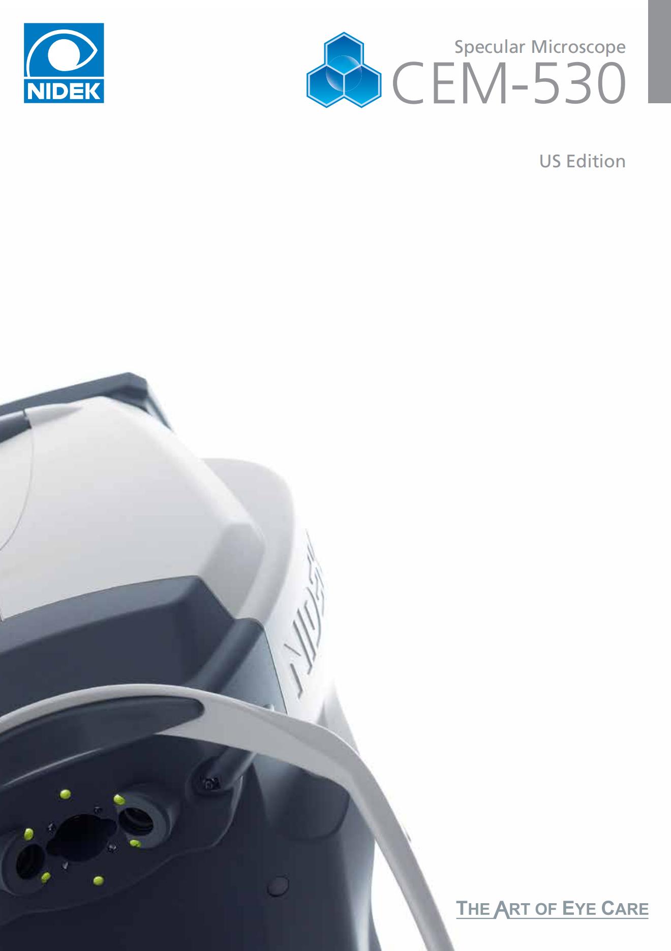
Specular microscopy
The specular microscope is a non-invasive imaging and analysis device of the corneal endothelial layer. The specular microscopy is an examination of endothelial cells using the specular reflexion resulting from the interface between endothelial cells and aqueous humour.
The different analysis zones
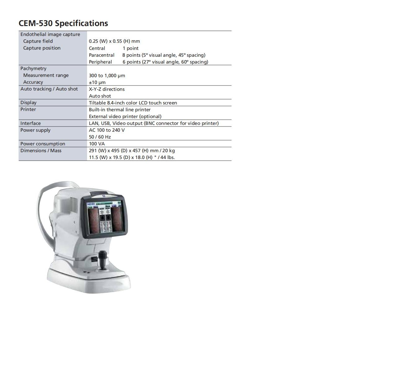
The specular microscope CEM-530 performs central, paracentral (diameter: 1.3 mm) and peripheral (diameter: 7.3 mm) conventional specular microscopies. The combination of central, paracentral and peripheral imaging provides an overview that can be used to make a detailed morphological and quantitative evaluation of the endothelial layer.
Paracentral images are captured from 8 points under a visual angle of 5° on a diameter of 1.3 mm to perform an improved evaluation surrounding the central appearance of the cornea.
Peripheral images are captured from 6 points under a visual angle of 27° on a diameter of 7.3 mm to perform an improved evaluation of the peripheral zone of the cornea.
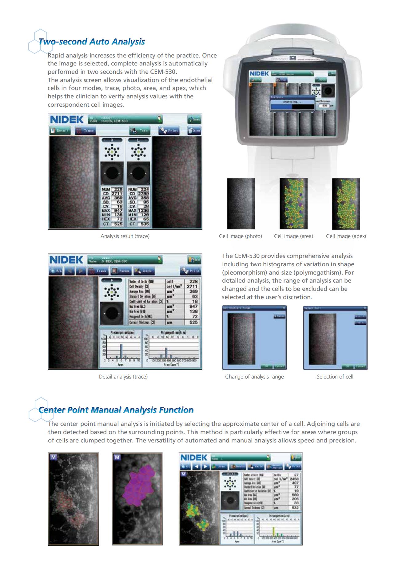
Quantitative & qualitative analysis
A series of 16 images are captured. These images are automatically sorted out depending on their quality. The built-in software identifies the image used to optimise the counting and perform the analysis of endothelial cells. Using the data is simple and quick.
Analysing the endothelium specular image consists in evaluating the appearance of cells covering this specific cornea layer to bring out anomalies such as drops or keratic precipitates. Very often, the operator has to count the number of cells in a defined area while analysing the distribution of the cell sizes.
Suitable for all practices
To perform a detailed analysis, the user can mark out the observation zone and take out of the counting the parts of the images that are inappropriate.
Moreover, a measurement of the central corneal thickness completes the data captured by the microscope.
In order to adapt to types of working organisation, connecting the CEM-530 to a network is possible. So it makes possible directly triggering from the machine the printing to a printer connected to the computer or directly sending the digital reports to a patient management software.
Viewer CEM-530 is available (optional) through the NAVIS-EX imaging software (patient data management software). The viewer CEM-530 allows a following-up of the counting of endothelium cells over time.